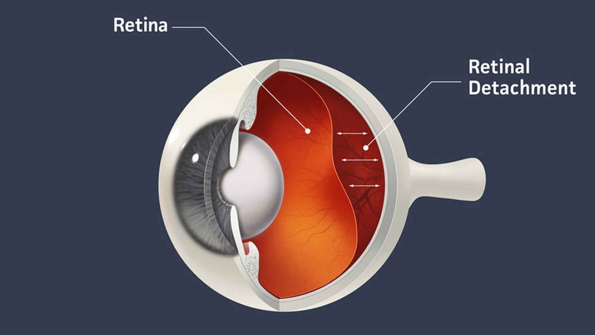
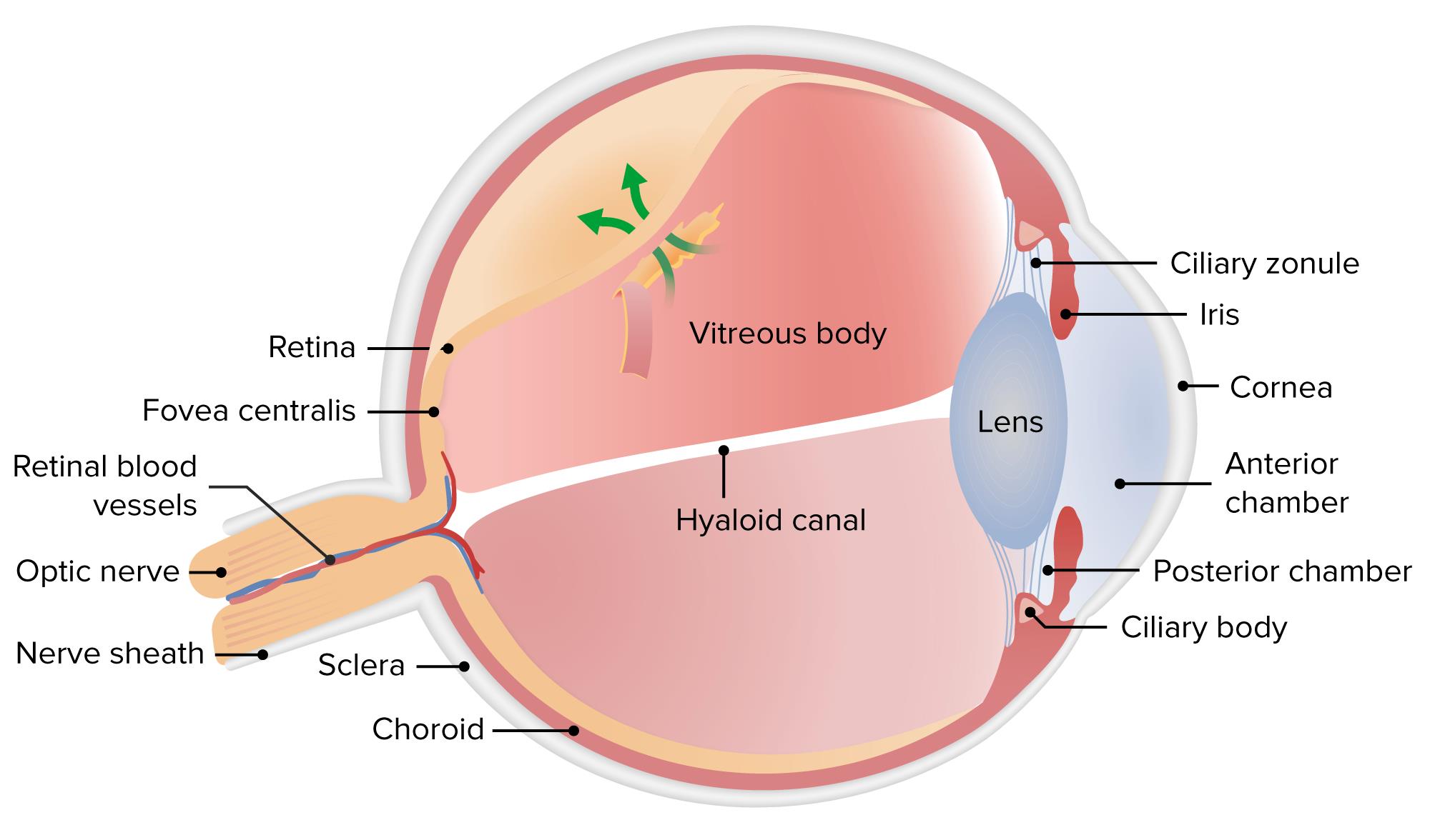
Retinal detachment is a serious eye condition in which the retina, the light-sensitive layer at the back of the eye, pulls away from its normal position. This separation prevents the retina from functioning properly, leading to vision impairment or blindness if left untreated. Common causes include aging, trauma, or underlying conditions like severe myopia. Early detection and prompt treatment are crucial to preventing permanent vision loss.

Retinal Detachment Symptoms
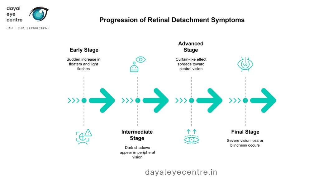
Recognizing the early warning signs of retinal detachment is crucial for timely medical intervention. Below are some of the key indicators:
Flashes of Light (Photopsia)
One of the most common symptoms of retinal detachment is experiencing brief flashes of light, known as photopsia. These flashes typically occur in the extreme peripheral vision and are unrelated to external light sources. They may appear as sudden, bright flickers, resembling lightning streaks. While occasional flashes can be harmless, persistent or increasing occurrences may signal a retinal tear or detachment.
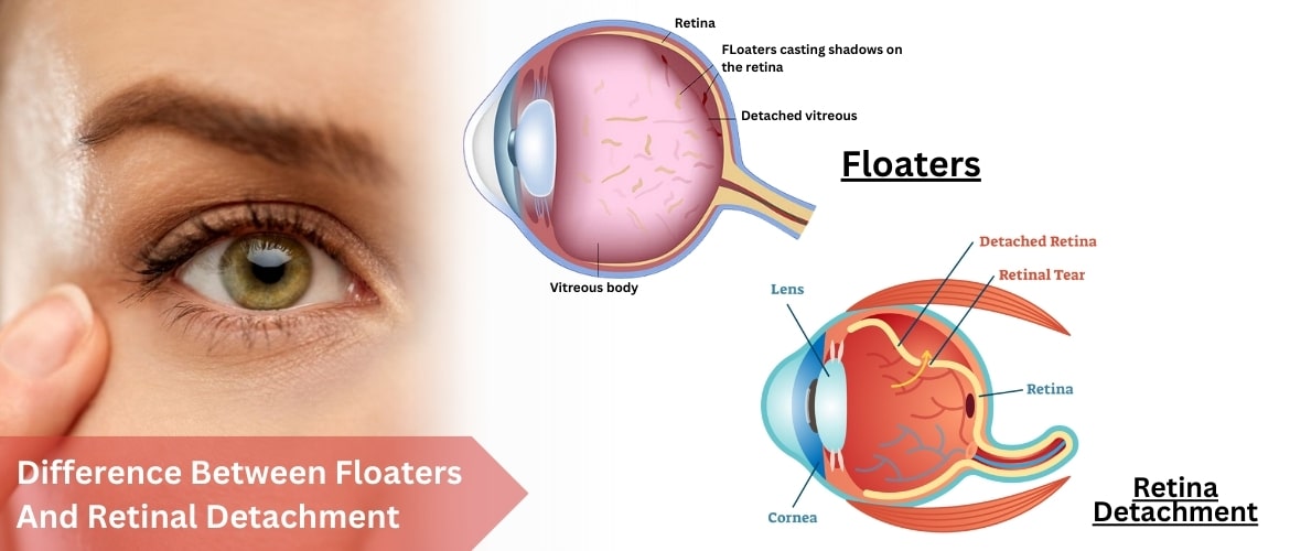
Sudden Increase in Floaters
Floaters are small, shadowy specks or thread-like shapes that move across your field of vision. While floaters are common with aging, a sudden and dramatic increase in their number could indicate retinal detachment. This happens when the vitreous gel inside the eye pulls away from the retina, sometimes leading to tears or breaks. Seeking immediate medical attention is recommended if new floaters appear alongside flashes of light.
Ring of Floaters Near Central Vision
In some cases, people experience a concentration of floaters forming a ring-like pattern near the temporal side of their central vision. This symptom may be an early warning sign of a retinal tear before full detachment occurs. If left untreated, it can progress to severe vision impairment.
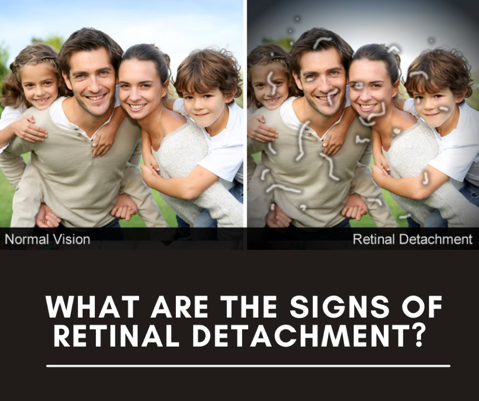
Curtain or Shadow Over Vision
A significant symptom of retinal detachment is the appearance of a dark shadow or curtain descending over part of the visual field. This shadow may begin at the sides (peripheral vision) and gradually move toward the center, obstructing sight. It may also feel as though a veil is covering parts of the vision. This symptom usually indicates a progressing retinal detachment, requiring immediate medical intervention.
Veil-Like Effect in the Field of Vision
Another striking symptom is the impression of a veil or curtain being drawn over the field of vision. This may occur suddenly or progress gradually, worsening over time. The severity depends on how much of the retina is affected. If ignored, this can lead to irreversible blindness.
Distorted Vision (Metamorphopsia)
Retinal detachment can cause straight lines to appear bent, wavy, or distorted. This distortion occurs due to the retinal layers shifting from their normal position, affecting how light is processed. People may struggle to read, recognize faces, or see fine details. If macular involvement occurs, distortion may be severe and permanent if left untreated.
Loss of Central Vision
As retinal detachment progresses, central vision may become blurry or disappear entirely. This happens when detachment spreads toward the macula, the part of the retina responsible for sharp, detailed vision. The extent of vision loss depends on the severity and duration of detachment. If the macula becomes fully detached, surgery must be performed urgently to restore vision, though full recovery may not always be possible.
Causes of Retinal Detachment
Retinal detachment can occur due to various underlying conditions and risk factors. The most common retinal detachment causes include severe myopia (nearsightedness), ocular trauma, previous eye surgeries, and diabetic retinopathy. Below are some of the major causes:
- Myopia (Nearsightedness) and Retinal Detachment: Severe myopia is a significant risk factor for retinal detachment. In individuals with high myopia, the eyeball is elongated, stretching the retina and making it thinner and more fragile. This increases the likelihood of retinal tears and lattice retinal degeneration, which can lead to retinal separation. Regular eye checkups are crucial for people with high myopia to monitor retinal health.
- Previous Cataract Surgery and Retinal Detachment: People who have undergone cataract surgery may have an increased risk of retinal detachment. During surgery, the natural lens is removed and replaced with an artificial intraocular lens (IOL). In some cases, this process can cause vitreous detachment, leading to retinal tears or exudative retinal detachment due to fluid accumulation. Patients who experience sudden retinal detachment eye flashes or floaters after cataract surgery should seek immediate medical attention.
- Ocular Trauma and Retinal Separation: Ocular trauma, including sports injuries, blunt force impacts, or accidents, can result in retinal detachment. A direct blow to the eye can cause the retina to tear or detach completely. Athletes and individuals in high-risk professions should use protective eyewear to minimize the chances of traumatic retinal detachment.
- Lattice Retinal Degeneration and Retinal Detachment: Lattice retinal degeneration is a condition where the peripheral retina becomes thinner and more vulnerable to tears. This degeneration is common in individuals with high myopia and can lead to spontaneous retinal detachment. Regular eye exams, including retinal detachment vision simulator tests, can help detect early signs of lattice degeneration and prevent serious complications.
- A Family History of Retinal Detachment: Genetics play a role in retinal detachment causes, as individuals with a family history of the condition are at higher risk. Certain inherited conditions, such as Stickler syndrome or Marfan syndrome, weaken retinal structures, increasing the chances of retinal separation. If there is a history of retinal detachment in the family, routine screenings are recommended to monitor retinal health.
- Diabetic Retinopathy and Retinal Detachment: Diabetes-related eye conditions, such as diabetic retinopathy, can lead to tractional retinal detachment. In advanced cases, abnormal blood vessels and scar tissue form on the retina, pulling it away from the back of the eye. This type of retinal detachment progresses gradually and may cause distorted vision, dark shadows, or central vision loss. Managing blood sugar levels and undergoing regular diabetic eye screenings can help prevent retinal separation.
What are the Risk Factors of Retinal Detachment?
Several risk factors increase the likelihood of developing retinal detachment. While some people may have a genetic predisposition, others may develop it due to injuries or underlying conditions. Below are the key risk factors:
- History of Retinal Detachment in One Eye: People who have had retinal detachment in one eye are at a higher risk of developing it in the other eye. Regular monitoring and timely intervention can help prevent further complications.
- History of Eye Surgeries (e.g., Cataract Removal): Individuals who have undergone cataract surgery or other intraocular procedures are more susceptible to retinal detachment. Surgical interventions can sometimes lead to vitreous detachment, increasing the chances of a retinal tear.
- Aging as a Risk Factor: Age-related changes in the vitreous gel inside the eye can contribute to retinal separation. As people age, the vitreous shrinks and may pull away from the retina, causing tears that lead to detachment. The risk is significantly higher after the age of 50.
- Severe Eye Injury and Retinal Detachment: Blunt trauma or penetrating injuries to the eye can cause retinal detachment by tearing the retina. Individuals involved in contact sports, high-impact activities, or accidents should take precautions to protect their eyes.
- Family History of Retinal Detachment: Genetics play a role in retinal detachment causes. If a close family member has experienced retinal detachment, the likelihood of developing the condition is higher. Routine eye checkups are essential for early detection.
- Myopia (Nearsightedness) and Retinal Detachment: People with high myopia (extreme nearsightedness) have elongated eyeballs, which stretch and thin the retina. This makes them more prone to lattice retinal degeneration and spontaneous retinal detachment.
- Underlying Eye Disorders and Diseases: Individuals with pre-existing eye conditions such as uveitis, lattice degeneration, retinoschisis, or Coats' disease are at greater risk of retinal detachment. These diseases weaken retinal structures, making them more vulnerable to separation.
Retinal Detachment Prevention
Preventing retinal detachment is crucial, especially for individuals at higher risk due to factors like myopia, previous eye surgeries, or systemic conditions like diabetes. While not all cases of retinal separation can be avoided, the following preventive measures can help reduce the risk:
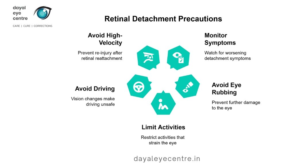
Avoid Direct and Indirect Eye Injuries
Eye trauma is a significant cause of retinal detachment, especially in people engaged in contact sports, hazardous jobs, or high-impact activities. To minimize the risk:
- Wear protective eyewear while playing sports like boxing, basketball, or racquetball.
- Use safety goggles when working in environments with flying debris or chemicals.
- Avoid rubbing the eyes aggressively, as excessive force can strain the retina.
- After an eye injury, seek immediate medical evaluation to rule out retinal tears or detachment.
Regular Eye Checkups
Routine eye examinations are vital for detecting early signs of retinal detachment, especially in individuals with high myopia, family history of retinal detachment, or pre-existing retinal conditions like lattice degeneration.
- People over 40 or those with risk factors should have an annual dilated eye exam.
- If experiencing retinal detachment eye flashes, sudden floaters, or vision distortions, consult an ophthalmologist immediately.
- Retinal detachment vision simulator tests can help patients understand potential vision changes and take preventive action.
Controlling Systemic Risk Factors and Diseases (e.g., Diabetes)
Systemic conditions such as diabetes and high blood pressure increase the likelihood of tractional retinal detachment due to abnormal blood vessel growth. To minimize risk:
- Maintain optimal blood sugar levels to prevent diabetic retinopathy.
- Manage high blood pressure and cholesterol through lifestyle modifications.
- Follow a healthy diet rich in antioxidants and omega-3 fatty acids, which support retinal health.